Fig 3a
Fig 3a – Intra-oral pictures of the patient. In these images we can see the partial edentulism in mandible and total edentulism in the maxillae. A) front view
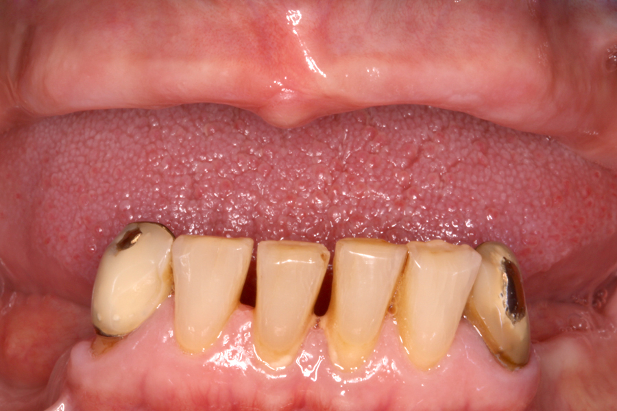
Fig 3a – Intra-oral pictures of the patient. In these images we can see the partial edentulism in mandible and total edentulism in the maxillae. A) front view