Dental implants in the aesthetic zone
An increasingly popular treatment is not without its risks. Full understanding of the process and potential complications will lead to a satisfied patient
Dr Michael Koukoulis
Dental implants are, with little doubt, the preferred choice for replacing missing or failing teeth. They are becoming increasingly popular, more widely available and more affordable as a treatment option.
It is therefore essential that the general dental practitioner has the necessary knowledge and tools to be able to assess and discuss in relative detail the option of dental implants with their patients, regardless of whether they are surgically placing them or restoring them.
The purpose of this article is to provide a quick overview of the specific challenges when considering dental implants in the aesthetic zone and the different approaches to meet these.
The Swiss Society of Oral Implantology developed the SAC classification system (Simple – Advanced – Complex) as a way to broadly categorise the difficulty of dental implant treatment. By that system, “a single tooth gap in the anterior maxilla without a bone defect present” is automatically classified as an Advanced case as it involves an area of high aesthetic demand combined with difficult pre-existing anatomy that presents both surgical and prosthetic challenges.
Clinical conditions that present with tissue deficiencies can be divided into two categories1.
1. Anatomic, which exist naturally, such as a narrow alveolar crest or the facial undercut of the alveolar process.
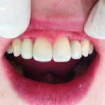
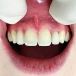
2. Pathologic, which includes dental trauma, acute or chronic infections (periodontal, periapical, endo-perio lesions) and bone atrophy due to long-term tooth loss. (Figures 1a, 1b, 2)
Faced with the above, the treating dentist/surgeon has to keep in mind the following surgical aspects for maximising the aesthetic outcome2.
- Pre-surgical planning
- Implant positioning
- “Aesthetic” bone grafting
- Soft tissue management.
1. Pre-surgical planning
Anatomic site analysis – This is arguably the most important decision-making step as it will determine the overall approach to the treatment. The general practitioner, even if not directly involved with implants, needs to be able to provide an initial assessment and have the confidence to discuss available options with the patient before proceeding with treatment or referring as required.
The alveolar crest needs to be evaluated intra-orally and radiographically. Is there a horizontal or vertical bone deficiency? Will a bone augmentation procedure be necessary for functional (i.e to maintain primary stability and long-term integration of the fixture) or aesthetic reasons (see below). To quote a colleague of mine, when assessing the aesthetic outcome of an implant, “soft tissue is the issue but bone sets the tone”. Hence there are
two critical bone structures under consideration:
The height and thickness of the facial/buccal bone wall
Several studies have shown that the concept of biologic width can be applied to osseointegrated implants3-5, in a similar manner that exists around natural teeth.
This translates to a relatively constant thickness of peri-implant soft tissues ofapproximately 3mm. Therefore, when a facial bone defectis present and in the absence of a bone augmentation procedure, one can expect soft tissue recession and an apically positioned gingival margin resulting in either exposure of the implant collar or an elongated crown with poor aesthetic outcome (Fig 3).
The height of the alveolar crest in interproximal areas
This effectively determines the presence or absence of peri-implant papillae (“black triangle disease”). It has been described as highly predictable in single-tooth gaps as it is dependent on the proximal bone level of the adjacent teeth and not the implant itself 6,7.
A distance of ≤6mm from the height of the crest to the contact point of the restoration will result in an increased chance of intact papillae filling in the interproximal spaces (Figs 4a, 4b). Inter-implant papillae in edentulous spaces of two or more missing teeth are not predictable, so it is important to discuss the aesthetic limitations prior to therapy to avoid unrealistic expectations. In these cases of multiple missing teeth, the use of implant supported bridges with ovate pontics to create pseudopapillae or the use of pink porcelain may be indicated.
In addition to the above, CBCT scans offer invaluable information with relation to the three-dimensional proximity of anatomical structures in the area such as the nasal floor and the nasopalatine canal, the position and axis of adjacent roots and any “foreign” bodies such as root filling materials, root fragments, unerupted teeth etc.
When assessing the implant site, it is important to avoid “tunnel vision” in concentrating on just the missing/failing tooth. The status of the adjacent dentition has to be evaluated
in terms of endodontic and periodontal health as well as structural crown integrity.
More often than not, for an anterior tooth to fail or be missing, there is a high chance that the adjacent teeth will be affected by some form of pathology. Their prognosis needs to be assessed and a decision to be made as to whether they can be treated or should be included in the overall implant treatment plan. In many cases, either of the above options is clinically acceptable so it is imperative to explain both to the patient and include them in the decision-making.
The dimensions of the edentulous space need to be measured to assess the potential aesthetic result and symmetry to the contralateral tooth. Even though size differences can be acceptable when replacing missing molars or even premolars, this is rarely the case with the upper anteriors. Therefore, the options of pre-operative orthodontics, enameloplasty, use of restorative materials or accepting a diastema need to be considered and discussed with the patient.
Looking at the soft tissues, two types of gingival morphotype (tissue biotype) have been described: a) Thick flat tissue with shallow scalloped gingivae, dense fibrotic tissue and a wide band of keratinized mucosa. This type is more resistant to recession and tends to respond with pocket formation following surgery or inflammation, making it more favourable when considering the aesthetics of peri-implant tissues. b) Thin, highly scalloped periodontium with a distinct disparity between facial and interproximal gingival levels. This type is more prone to recession therefore in such cases, a further palatal and deeper placement of the implant shoulder should be considered to mask potential show-through and create a smoother emergence profile of the restoration.
Finally, the patient’s lip and smile line (high – medium – low) need to be assessed as it can have dramatic implications on the overall aesthetic outcome and will ultimately guide our approach to treatment.
Once the implant site has been assessed, a decision needs to be made with regards to the type of surgical protocol to be followed. Broadly speaking there are three distinct options:
Delayed placement – where the tooth/teeth have been missing for more than six months. These cases can exhibit variable degrees of bone resorption but benefit from healed sites with mature hard and soft tissues and the resolution (usually!) of any pre-existing pathology.
Immediate placement – where the implant fixture is placed directly into the socket of the tooth at time of extraction. Literature has shown high success rates with this approach and has a strong appeal to patients with the clear advantage of reduced treatment times and fewer surgical appointments. It has also been suggested that by immediately inserting the implant, which acts as a tooth root, it may lead to preservation of the hard and soft tissues following extraction. On the other hand, there is potentially a higher risk of complications or a compromised aesthetic result due to a combination of:
- Pre-existing pathology/acute infection
- Difficulty to achieve the ideal three-dimensional positioning of the implant (see below)
- Traumatic manipulation of soft issues at time of extraction
- Increased osteoclastic activity following the removal of the tooth
- Challenging soft tissue closure over the socket.
- Para-immediate placement – where the implant is placed six to eight weeks following extraction. This approach allows for a soft tissue seal to develop, high osteoclastic activity to reduce and also time for any acute infection and inflammation to resolve. Nevertheless, a simultaneous bone-grafting procedure will likely be required at time of implant placement.
2. Implant positioning
The correct three-dimensional positioning of the implant shoulder is key in achieving
an aesthetic and harmonious outcome. Surgical placement of the implant needs to be restorative-driven, i.e work backwards from the proposed restoration. This is achieved with the use of a preoperative wax-up (or the use of an existing tooth/crown if satisfactory) to determine the facial profile and gingival margin of the final restoration.
A simple clear surgical template can then be fabricated that highlights the position of the buccal gingival margin and will guide the placement to achieve the ideal emergence profile and long-term peri-implant hard and soft tissue support.
The implant shoulder position can be viewed in the following three dimensions: Mesiodistal, Orofacial (labiopalatal) and Apicocoronal. “Comfort” and “danger” zones can then be defined within these dimensions in relation to achieving an aesthetic outcome.
Mesiodistal – danger zones are located next to adjacent teeth. Literature varies but general consensus is that a minimal distance of 1-1.5mm between implant and root surface and ~3mm between two implants should be maintained. Encroaching on that space can result in resorption of the interproximal alveolar crest with reduction/loss of the interproximal papillae and an asymmetrical restoration with poor embrasure form and long contact zone.
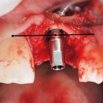
Orofacial – the facial aspect of the implant shoulder should be approximately 1mm palatal to the point of emergence of the proposed restoration or that of the adjacent teeth (Fig 5) 8. Too far facially and we see buccal bone loss with soft tissue recession. Too far palatal and then there are issues with the emergence profile resulting in a ridge-lap restoration, which can be un-aesthetic and difficult to maintain.
Apicocoronal – “comfort” zone for the implant shoulder is about 1mm apical to the CEJ
of the adjacent teeth (if no existing gingival recession is present!). More realistically, the same comfort zone is approximately 2mm apical to the midfacial margin of the planned restoration (Fig 6). The principle of “as shallow as possible, as deep as necessary” can be kept in mind in an effort to balance aesthetic and biologic principles. If the implant is placed too deep, it will result in undesired facial and interproximal bone loss as well as prosthetic difficulties. Too shallow of a placement can lead to visible metal margins, poor emergence profile and “square” crown morphology.
3. Aesthetic bone grafting
Can be defined as the regeneration of osseous foundation to serve aesthetic purposes (such as soft tissue support, ideal implant positioning etc.) where it is not “essential” to the stability and osseointegration of the fixture itself. The objective is to augment bone mainly in the horizontal direction and establish a thick facial bone wall for long-lasting soft tissue support. The GBR technique shows good results throughout the literature for this. Hard and soft tissue grafting in the vertical dimension appears to be more complex with less predictable results, and it is important to discuss this with the patient before the onset of treatment.
4. Soft tissue management
Pre-surgical: This includes techniques aiming to increase existing keratinised tissue such as root reduction prior to extraction followed by immediate implant placement.
At time of surgery: The objective is to minimise traumatic manipulation of the soft
tissues. Parapillary incisions are preferable where possible as they have shown to reduce risk of recession/loss of the interdental papillae. Tension-free flap closure is paramount to maintain blood supply, especially in cases where a bone augmentation procedure is carried out. This is achieved by extending vertical incisions and deep periosteal slitting to freely mobilise the flap. Flapless surgery, if indicated, offers the most conservative approach with regards to the soft tissues, but detailed three-dimesional imaging +/- surgical guide for accurate positioning of implant may be necessary.
At abutment connection: The modified roll flap is a relatively simple technique to try to increase soft tissue height labially. It is carried out in cases of a two-stage healing protocol when the implant is uncovered and a healing cover or abutment + provisional restoration are fitted.
Alternatively, conventional connective tissue grafts can be also performed at this stage. From the prosthetic point of view, the use of ovate pontics as provisionals can help to create
an emergence profile that imitates that of the natural tooth and enhance formation of “pseudopapillae” between adjacent implants. The use of custom- milled healing abutments has also been described. These are used in sequence from smaller to larger to gradually expand the tissues and increase the band of keratinised gingivae. Finally, custom-milled zirconia abutments can offer more natural aesthetics in combination with all ceramic crowns and reduce the risk of metal show-through in case of thin gingivae coronally.
Discussion
There is a multitude of factors to consider when planning or placing dental implant restorations in the aesthetic zone, which are well beyond the scope of this article. New materials (implant surfaces, bone and soft tissue grafts) are developed continuously with new protocols to surgical place and restore implants. In an ideal world with unlimited time and resources, every single implant case would undergo bone and soft tissue grafting with long-term provisional restorations to shape and achieve the best aesthetic outcome. In reality though, aside from the clinical factors mentioned above, the condition of the remaining dentition, finances, treatment duration, need for provisional restorations etc. play an equal role in the decision process.
Therefore it is essential to assess overall patient expectations, explain the treatment options available along with potential challenges/complications and make a decision with the patient on how to approach their treatment.
Full understanding of the process, outcomes and potential complications will lead to a satisfied patient as much as sound clinical skills.
Dr Michael Koukoulis, B.Dent.S (Hons) (Glasgow 2003), Pg.Cert Implant Dentistry (Warwick 2006)
References
1. Buser D, Martin W, Belser UC. Optimizing Esthetics for Implant Restorations in the Anterior Maxilla: Anatomic and Surgical Considerations. (Int J Oral Max Implants 2004;19:43-61)
2. El Askary AS. Multifaceted Aspects of Implant Esthetics: The Anterior Maxilla. (Implant Dent 2001; 10: 182-191)
3. Berglundh T, Lindhe J. Dimension of the Periimplant Mucosa. Biological width revisited. (J Clin Periodontology 1996; 23: 971-973)
4. Cochran DL, Hermann JS, Schenk RK, Higginbottom FL, Buser D. Biologic Width around Titanium Impalnts. A histometric analysis of the implanto-gingival junction. (J Periodontology 1997; 68: 186-198)
5. Hermann JS, Buser D, Schenk RK, Higginbottom FL, Cochran DL. Biologic width around titanium implants. A physiologically formed and stable dimension over time. (Clin Oral Implant Res 2000; 11: 1-11)
6. Kan JY, Rungcharassaeng K, Umezu K, Kois JC. Dimensionsof peri-implant mucosa: An evaluation of maxillary anterior single implants in humans (J Periodontology 2003; 4: 557-562)
7. Salama H, Salama M, Garber D et al. The interproximal height of bone a guide post to predictable esthetic strategies and soft tissue contours in anterior tooth replacement. (Pract Periodontics Aesthet Dent 1998; 10: 1131-1141)
8. Belser UC, Bernard JP, Buser D. Implant supported restorations in the anterior region: Prosthetic considerations. (Pract Periodontics Aesthet Dent 1996; 8: 875-883)
Fig. References
-
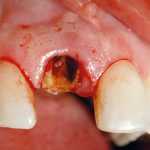
- Figure 1a: Typical loss of buccal bone plate folloing root fracture of UR1 that was previously restored with a post crown
-
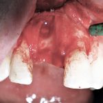
- Figure 1b: Typical loss of buccal bone plate folloing root fracture of UR1 that was previously restored with a post crown
-
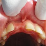
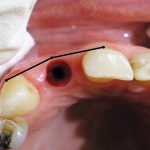
- Fig 2: Buccal bone atrophy due to long-term loss of UR1
-
- Fig 3: Recession of gingival margin around UL1 implant due to lack of adequate buccal bone support
-
- Fig 4a: UR2 and UL2 implant restorations at fit
-
- Fig 4b: UR2 and UL2 one-year post-op showing infill of interproximal papillae
-
- Fig 5: Orofacial positioning of implant shoulder showing facial aspect 1mm palatal to point of emergence of adjacent teeth
-
- Fig 6: Apicocoronal position showing implant shoulder 2mm apical to the midfacial margin of the planned restoration
Tags: 2018, aesthetic, aesthetic zone, Dental implants, Implant dentistry, interproximal papillae, July, July 2018, Koukoulis, Michael Koukoulis
You must be logged in to post a comment.