A real success story
James Hamill, BDS MFDs RCSEd, presents the case that won the Smile Award for Single Implant Smile at the 2010 awards ceremony in London
Presenting Situation
The patient, a 45-year-old female, attended following referral on 17/08/06 in regard to the possible replacement of the UR1 which was currently restored with an upper partial CCR denture. The patient expressed a wish to get rid of the existing unsightly denture and have this replaced with a ‘fixed’ tooth.
Relevant dental and social history
The patient had attended her own dentist for routine examination and requested the possibility of replacing her single tooth denture with a dental implant. She had been made aware of this possibility through her own research and was excited at the possibility this would offer her.
The UR1 was traumatically avulsed 10 years previously and had not seen a dentist since the denture was fitted following the accident. There had been virtually no dental intervention during the patient’s lifetime. The patient was medically fit and well but did have quite severe arthritis affecting her hands – this was making it more difficult to remove the denture on a routine basis.
Patient expectations and wishes for treatment
The patient wished to be rid of the denture, which was a constant reminder to her of the accident that led to the tooth loss 10 years earlier. Her main wish, if she was deemed suitable, was for a dental implant to be placed. She requested that the implant-retained crown would improve her appearance.
The patient had been brought up to speed by her referral dentist with regard to implant therapy and so felt that she was prepared for treatment.
Initial clinical findings
Extra oral:
Patient was deemed to have a low smile line, a pleasant appearance with a full perioral tissue appearance. There was some guarding of the smile.
• The UL1 rested on the lower lip at rest
• No TMJD was noted or reported
•With the denture in place the smile was affected.
Intra oral:
No caries were detected and oral hygiene was deemed excellent, with no periodontal issues seen. All pockets were
Initial implant assessment was carried out and an aesthetic risk assessment as per ITI guidelines was carried out. This included the below factors and how they influenced risk:
• Non smoker – low risk
• Medically fit and well – low risk. The only addition to this was the arthritis and possible cleaning issues in the future. This was deemed to be the same for any restoration used and so considered low risk
• Adjacent teeth healthy and unrestored – low risk
• Gingival biotype was thick and low scalloped – low risk, but increased risk of scarring and blunted papillae. In fact, here there was an excess of tissue so tissue contouring was anticipated. There was a strong frenum associated and it was anticipated that this may need to be relieved
• Tooth shape – triangular putting the risk factor to high for soft tissue aesthetics.
• Patients expectation – seen as medium
• Implant site – there was no infection hence low risk
• Bone level to adjacent teeth at implant site – medium risk
• Bone availability – there was a horizontal defect giving medium risk.
The occlusion was favourable, however the contralateral UL1 tooth shape and colour was deemed complex. The adjacent teeth were vital to EC and EPT testing.
Radiographs
PA of UR1 was taken. The PA revealed favourable adjacent root position for a dental implant.
Diagnosis
An avulsed UR1 due to trauma required replacement with a dental implant or other suitable restoration. This was to be considered in a well cared for mouth with excellent oral hygiene and periodontal status with an overall medium aesthetic risk profile as per the ITI guidelines.
A horizontal bone defect did exist, which would require augmentation if the implant route was to be followed. Excess and thick soft tissue present, leaving the need for accurate soft tissue management.
Initial treatment plan discussion
Following the initial examination a discussion was had with the patient about her treatment options. These were:
1) Orthodontics to close the space. This was immediately ruled out
2) A new denture. This would not have fulfilled the patient’s initial expectations
3) An adhesive bridge
4) A conventional bridge
5) A dental implant and crown
6) Accept current situation – ruled out.
The discussion revolved around the above options. Options one, two and six were quickly disregarded for obvious reasons. Option four was discussed but we both felt that the necessary tooth reduction was not acceptable and the restorative cycle that would ensue was not welcomed by the patient.
Option three was considered for some time. The main issues were going to be pontic design and ease of oral hygiene if the arthritis worsened, shine through of the wing material, the occlusion which left the need for tooth preparation and the issues of debonding. The cost was attractive to the patient at this stage as was the apparent speed of treatment.
Implants were then discussed. We advised that the bone would require grafting, the tissue would require considerable management in a provisional restorative phase and that the time and cost would be high. We discussed the challenging colour match and tooth morphology. The advantages of having a predictable fixed tooth result were discussed without the need to remove any adjacent tooth tissue.
A summary discussion was then had and the patient decided initially that an implant was the treatment of choice for her. This was to be confirmed by letter and patient agreement.
Final treatment plan and rationale
• Following a treatment plan letter outlining the options and cost implications the patient decided on the implant option. A patient agreement was signed and the treatment plan agreed.
• Impressions were taken for the fabrication of a surgical stent. This was designed for the placement of a Straumann 4.1mm diameter RN Tissue level implant. The implant was to be placed to allow for a cement-retained final restoration. The stent design incorporated the proposed gingival emergence of the final crown to aid 3D implant placement. Impressions also taken for the manufacture of a partial acrylic denture to wear during healing. This was to be tooth- supported with ball ended clasps in the URQ and ULQ.
• Implant placement under LA with horizontal simultaneous grafting using Bio-oss and Bio-gide – to ease soft tissue
• Sutures removal at 10 days
• Review of integration at four weeks
• Primary fixture head impression at eight to 10 weeks
• Fitting of a cement retained provisional crown
• Review of tissue progress
• Further review of progess
• Final tissue review
• Final fixture head impression using a modified technique
• Final fitting of the restoration using a CARES Zirconia abutment with an all ceramic cementable crown.
Treatment timescale
03/08/06 Initial consultation
07/09/06 Agreement of treatment plan
16/10/06 Implant placement.
This was as expected with a 4.1 RN SP 12mm implant placed. This was carefully sited in a 3D fashion using the stent as a guide. Horizontal augmentation was required using Bio-oss and double layer Bio-gide membrane. A frenotomy was carried out to help relieve tissue tension on primary closure over an internal healing abutment.
29/10/06 Suture removal.
21/11/06 Integration review at four weeks. Healing was seen to be excellent with an excess of vertical tissue. This was thick and dense. The implant was uncovered and a bevelled healing cap placed. The uncovering was with a 5mm tissue biopsy punch.
08/01/07 Primary fixture head impression taken for the manufacture of an acrylic cement retained provisional crown. A screw retained impression coping was used.
30/01/07 Fitting of the acrylic provisional crown. This was cemented with clear ‘temp bond’ and the blanching allowed five minutes to resolve. This happened and the patient was discharged.
30/03/07 Review of tissue maturation. Things were going well with a good mesial
papilla; the distal was blunted and round and was slowly filling from the palatal. Some composite was added to the distal aspect of the provisional restoration and again blanching was allowed to recover before the patient departed.
17/04/07 Review of tissue maturation. Some of the distal composite was removed as it was deemed on reflection that too much had been added. The mid face was recontoured to try to achieve the distinctive shaping on the adjacent UL1.
07/06/07 Review of tissue maturation. Tissues were responding well and the overall shape was doing nicely.
04/07/07 Review of tissue maturation. Eased distal aspect again to encourage distal papilla.
15/08/07 Final review and all agreed that we would finish.
16/10/07 Modified impression taken.
19/11/07 Final fit of UR1 crown. CARES abutment fitted and the final crown cemented in place. There was some small degree of blanching but this settled quickly.
20/12/07 Final review. Patient was delighted with the result. Team also delighted. Discharged back to referral dentist and 12-month review with the Blueapple team.
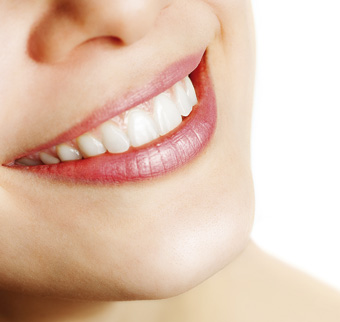
Final result
Following the gingival contouring with the temporary restoration, the patient was restored with an all-ceramic crown on a CARES zirconia abutment. The overall result was very pleasing considering the degree of soft tissue manipulation required. It was a credit to the patient for her patience and the technical control of the provisional phases of treatment. The final ceramic work was superbly carried out by the ceramist.
Total treatment time from the initial consultation to the fit of the final restorations was 16 months. A reminder that single implant cases in the anterior maxilla can never be classed as straightforward.
James Hamill, BDS MFDS RCSEd, is the principal dentist at Blueapple in Belcoo, County Fermanagh. He qualified from the University of Dundee in 2001 and gained his membership of the Fellowship of Dental Surgeons of the Royal College of Surgeons of Edinburgh in 2004. He is also the UK and Ireland speaker for the ITI (International Team for Implantology).
Patient testimonial:
“After losing my front tooth 10 years ago, I was delighted and excited to be referred to see James to find out if I could see the back of my denture by using a dental implant. Having read up on the subject I was surprised just what was involved in my case. James clearly told me that it would take time to get a good result and I was happy to trust him. Looking now at the final result, it was all really worth it, the tooth is indistinguishable from its neighbour and I am very, very happy with the final appearance. Thanks to all at Blueapple, I look forward to seeing you all soon.”